Choledochal Cyst
Learn About Choledochal Cysts and Dr. Sucandy Choledochal Cysts Treatments. Complete Form
 Choledochal cysts, are rare congenital dilations (enlargements) of the bile ducts, a biliary tube that drains bile produced by the liver into small intestine for food digestion. They are increasingly diagnosed in adults. Choledochal cysts are inflammatory in nature. Left untreated, choledochal cyst frequently leads to recurrent biliary infection, biliary stasis, or pancreatitis (inflammation of the pancreas) and ultimacalltoy causes biliary cancer from the chronic inflammatory state.
There are 5 types of choledochal cysts, based on the location of cyst within the biliary tree.
choledochal cyst types
Choledochal cyst is a premalignant condition with substantial risk of malignant transformation into cholangiocarcinoma, bile duct cancer. Cholangiocarcinoma is a type of highly aggressive cancer with a poor prognosis. Diagnosis of choledochal cyst is obtained with CT scan or MRI/MRCP scan. The entire biliary tract can be depicted in a high-quality MRI/MRCP scan. A cystic dilation of the biliary tree is consistent with the diagnosis of choledochal cyst. Identification of the surrounding structures such as hepatic artery and portal vein is also important for preoperative planning.
The standard treatment for choledochal cyst is via complete cyst resection with or without liver resection, depending on type of the choledochal cyst, followed by a reconstruction of the biliary tree. Choledochal cyst that involves liver parenchyma require a partial liver resection. Choledochal cyst excision/resection is best achieved through minimally invasive surgical technique. When compared to open operation, minimally invasive surgery leads to lower postoperative complication, less pain, less requirement for narcotics pain medication, and shorter recovery. Only very few centers in the United States perform minimally invasive choledochal cyst resection. A high-volume bile duct surgeon or biliary center achieves superior outcomes when compared to a low-volume bile duct surgeon or biliary center.
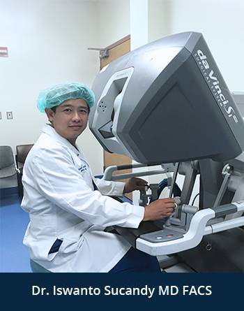
Dr. Iswanto Sucandy offers minimally invasive choledochal cyst resection/excision using the robotic technique. We have achieved significant experience in biliary tract surgery and liver surgery over the years. Our team work collaboratively with our interventional radiologists and interventional endoscopists to achieve the best long-term outcomes.


